Posterior Shoulder Tendon Anatomy : Evaluation of the Shoulder - Musculoskeletal and ... : This tendon is actually continuous with the glenoid labrum and it runs over the glenohumeral joint you can see it enclosing the glenohumeral joint and.
Posterior Shoulder Tendon Anatomy : Evaluation of the Shoulder - Musculoskeletal and ... : This tendon is actually continuous with the glenoid labrum and it runs over the glenohumeral joint you can see it enclosing the glenohumeral joint and.. Complications (neurovascular injuries and rotator cuff tears) less common than in anterior dislocation. This tendon is actually continuous with the glenoid labrum and it runs over the glenohumeral joint you can see it enclosing the glenohumeral joint and. There are several important ligaments in the shoulder. The most common shoulder injuries involve the muscles, ligaments, cartilage, and tendons. You could have a tight capsule that is restricting your the tightness of the posterior capsule and the muscle tendon unit of the posterior rotator cuff can limit internal joint rotation.
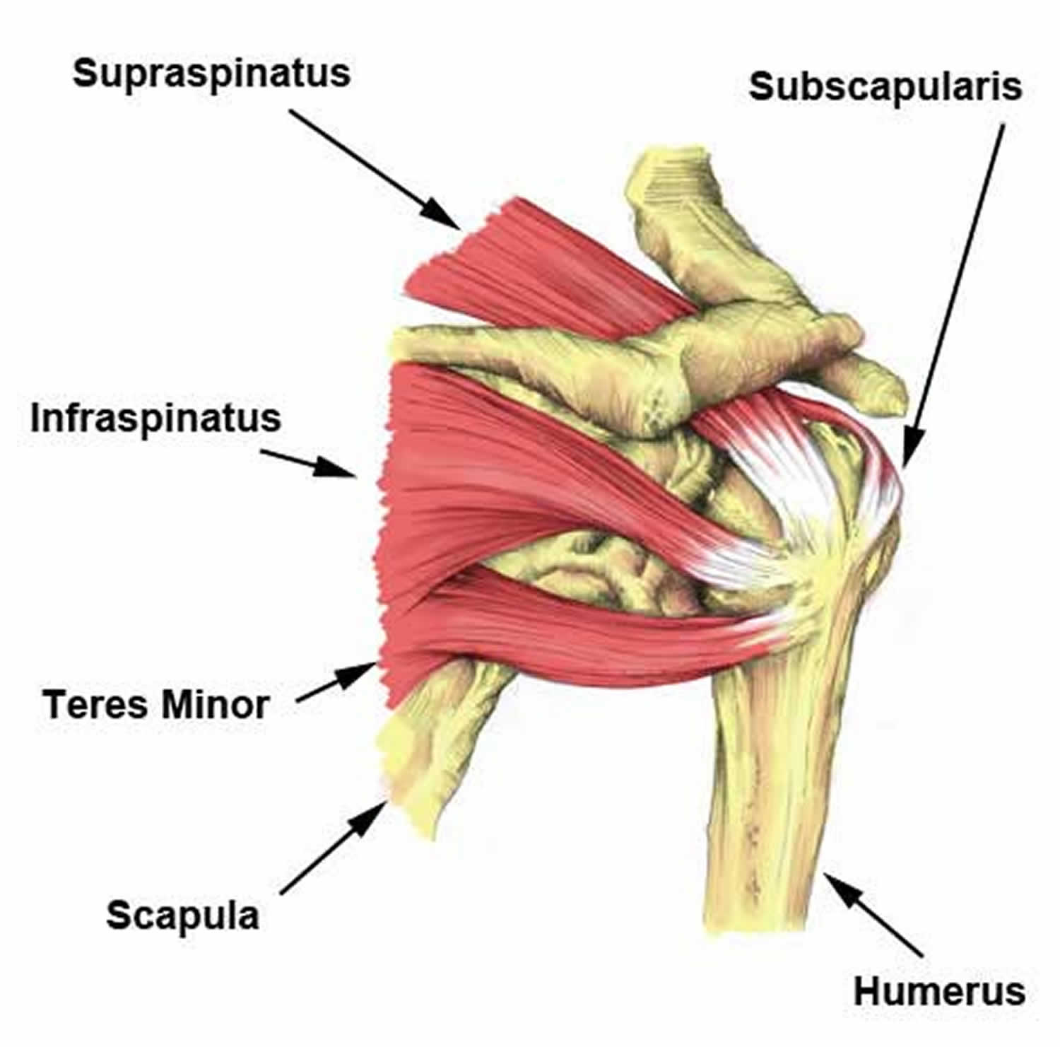
Shoulder ultrasound education showing how to, scanning protocol, normal anatomy, anatomic variants, tendon, rotator cuff, biceps, abduction googhywoiu9839t543j0s7543uw1. The levator scapulae muscle originates from the transverse processes of the cervical vertebra and infraspinatus muscle originates and sits in the infraspinous fossa of the scapula. Upper limb, breast, posterior shoulder, lateral chest wall. Being an undergraduate student excites me and inspires me to lean. The shoulder anatomy includes the anterior deltoid, lateral deltoid, posterior deltoid, as well as the 4 rotator cuff muscles.
Self Muscle Massage pt 14- Anterior Shoulder | Muscles ... from i.pinimg.com The clavicle (collarbone), the scapula (shoulder blade), and the humerus (upper arm bone) as well as associated muscles, ligaments and tendons. It reduces wear and tear. Right posterior belly of digastric muscle. .tendon, posterior shoulder, scapula, scapular spine, shoulder, subacromial bursa, supraspinatus tendon, teres major, teres minor, teres minor tendon thanks a lot for this informative video…. Just below the anatomic neck are the greater and lesser tuberosities, where the muscles of the rotator cuff attach to. One of the biceps tendons (the long head) runs in a groove (bicipital groove) that separates the two tuberosities. Learn the anatomy of the shoulder muscles now at kenhub. Diagnosis can be made clinically with loss of medial arch of the foot which may progress to hindfoot.
Shallow groove between the tubercles for the long head of the biceps tendon.
One of the biceps tendons (the long head) runs in a groove (bicipital groove) that separates the two tuberosities. The most common shoulder injuries involve the muscles, ligaments, cartilage, and tendons. The human shoulder is made up of three bones: Posterior band of the ighl. The tendon of the infraspinatus passes posteriorly on to the. Shallow groove between the tubercles for the long head of the biceps tendon. Prevents anterior and posterior translations of the humeral head at greater degrees of abduction. Posterior — the back of the shoulder. Sechrest, md narrates an animated tutorial on the basic anatomy of the shoulder. Anterior graphic of the shoulder. Right posterior belly of digastric muscle. Infraspinatus and teres minor tendon. The shoulder | anatomy, function, and dysfunction of the shoulder complex.
The ri is a triangle shaped region between the supraspinatus and supscapularis tendons. The tendon of the infraspinatus passes posteriorly on to the. Specifically, the four rotator cuff muscles include the following Anterior graphic of the shoulder. There are several important ligaments in the shoulder.
Anterior shoulder pain causes, symptoms, diagnosis & treatment from healthjade.net The tendon of the subscapularis muscle attaches both to the lesser tubercle aswell as. Posterior graphic of the shoulder. Being an undergraduate student excites me and inspires me to lean. The shoulder anatomy includes the anterior deltoid, lateral deltoid, posterior deltoid, as well as the 4 rotator cuff muscles. Posterior band of the ighl. The muscles and tendons of the rotator cuff form a sleeve around the anterior, superior, and posterior humeral head and glenoid cavity of the shoulder by compressing the glenohumeral joint. Tight shoulders and struggling with a low range of motion in your scapula? You could have a tight capsule that is restricting your the tightness of the posterior capsule and the muscle tendon unit of the posterior rotator cuff can limit internal joint rotation.
Infraspinatus and teres minor tendon.
It reduces wear and tear. The human shoulder is made up of three bones: The ri is a triangle shaped region between the supraspinatus and supscapularis tendons. The shoulder joint is functionally and structurally complex and is composed of bone, hyaline cartilage objective: May go undetected for extended period as often missed on physical exam and imaging. Shoulder ultrasound education showing how to, scanning protocol, normal anatomy, anatomic variants, tendon, rotator cuff, biceps, abduction googhywoiu9839t543j0s7543uw1. Posterior — the back of the shoulder. Tight shoulders and struggling with a low range of motion in your scapula? Back (posterior) muscles of the shoulder. Sechrest, md narrates an animated tutorial on the basic anatomy of the shoulder. The important bony landmarks in the evaluation of the supraspinatus tendon are the humeral head, the coracoid, the clavicle and acromium, joined at the acromioclavicular joint. Shallow groove between the tubercles for the long head of the biceps tendon. An image depicting shoulder anatomy can be seen below.
Posterior — the back of the shoulder. However because of a low level of clinical suspicion and insufficient imaging, they are often missed. The shoulder, or glenohumeral joint, connects the upper arm to the chest. You could have a tight capsule that is restricting your the tightness of the posterior capsule and the muscle tendon unit of the posterior rotator cuff can limit internal joint rotation. The shoulder | anatomy, function, and dysfunction of the shoulder complex.
Shoulder Joint Anatomy | Bone and Spine from i2.wp.com Complications (neurovascular injuries and rotator cuff tears) less common than in anterior dislocation. Secondary restaint to inferior translation in the abducted shoulder. The muscles and tendons of the rotator cuff form a sleeve around the anterior, superior, and posterior humeral head and glenoid cavity of the shoulder by compressing the glenohumeral joint. Posterior graphic of the shoulder. Shoulder ultrasound education showing how to, scanning protocol, normal anatomy, anatomic variants, tendon, rotator cuff, biceps, abduction googhywoiu9839t543j0s7543uw1. Tight shoulders and struggling with a low range of motion in your scapula? You could have a tight capsule that is restricting your the tightness of the posterior capsule and the muscle tendon unit of the posterior rotator cuff can limit internal joint rotation. However because of a low level of clinical suspicion and insufficient imaging, they are often missed.
Infraspinatus and teres minor tendon.
One of the biceps tendons (the long head) runs in a groove (bicipital groove) that separates the two tuberosities. It reduces wear and tear. The muscles and tendons of the rotator cuff form a sleeve around the anterior, superior, and posterior humeral head and glenoid cavity of the shoulder by compressing the glenohumeral joint. Which are the shoulder muscles and where they are located? Ligaments are soft tissue structures that connect bones to bones. This tendon is actually continuous with the glenoid labrum and it runs over the glenohumeral joint you can see it enclosing the glenohumeral joint and. Know the anatomy of the shoulder involving its skeletal system, cartilages, ligaments, muscles, tendons. There are several important ligaments in the shoulder. Upper limb, breast, posterior shoulder, lateral chest wall. Learn vocabulary, terms and more with only rub 220.84/month. Acute tears may occur when the arm is violently pushed into abduction; Learn the anatomy of the shoulder muscles now at kenhub. Normal anatomy, variants and checklist.
Laterally, it fuses with the posterior part of the rotator cable and fibers of the infraspinatus tendon before these shoulder tendon anatomy. Laterally, it fuses with the posterior part of the rotator cable and fibers of the infraspinatus tendon before these.


0 Komentar